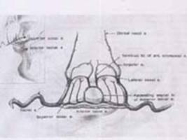
Normal Anatomy of lip and palate
The ultimate goal in treating cleft patients is to perform reconstructive surgery that creates a normal appearance and restores function. The following will describe the normal anatomy of the lip, nose and palate.
The Upper lip
Vermilion: The lower margin of the upper lip is called the vermilion and is characterized by its rosy color. The line or ridge between the skin of the upper lip and the vermilion is called the vermilion border.
Cupid’s bow: Is used to describe the concave or dipped portion of the vermilion border in the center of the upper lip.
Philtrum: Above the center of the upper lip is a dimple called the philtral dimple, and the raised ridges on either side of this dimple are the philtral columns or lines. The portion of the upper lip between the two philtral columns is known as the philtrums. In unilateral clefts, the philtrum remain attached to the larger portion of the lip. In bilateral clefts, the philtrum is isolated from the lateral lip segments. In this case, it is called the prolabium.
The Upper lip is composed of orbicularis oris muscle covered by skin on the outside and mucous membrane on the inside. Many other muscles are attached to the orbicularis oris muscle, and together they work to create the movements and power required for speaking, eating and forming facial expression. The orbicularis oris muscle constitutes the major bulk of the upper and lower lips.


The Nose
The nose can be thought of as a tent of skin supported in the center by a post of cartilage called the nasal septum which separates the nose into the right and left chambers. The nasal septum also separates the two large sinuses underlying the cheek bones which are called the maxillary sinuses.The shelves on the lateral walls of the nasal cavities are called turbinates. The nostrils include the external ports or openings as well as the skin and cartilage surrounding them. In unilateral and bilateral cleft, the nostrils are deformed.


The Palate
The palate is the roof of the mouth. It consists of two portions the hard (bony) palate and the soft, muscular palate. The palate separates the nasal cavity from the oral cavity.
The hard palate is a composite of several bony structures. The arch of the hard palate is called the palatal arch.
The hard palate constitutes the anterior portion of the entire palate and lies directly behind a horseshoe-shaped bony arch which supports the teeth. This arch is called the alveolus. The teeth protrude from a ridge known as the alveolar ridge. The hard palate is immobile. The flesh that covers the hard palate is called mucoperiosteum. It is used in cleft palate repair to close the hard palate defect.
The soft palate lies behind the hard palate. It ends with a little flap that hangs down from the soft palate called the uvula. The soft palate is composed of several muscles and fibrous tissue (all of which is attached to the posterior edge of the bony palate). The soft palate is mobile and plays a decisive role in speech production. This primary function of the soft palate is dependent on the levator (veli palatine) muscle. Reconstruction of this muscle is an important part of cleft palate surgery.
Behind the palate lies the pharynx or throat. The pharynx begins behind the mouth is called the oropharynx.
The soft palate performs many functions. One of its roles is to close off the back of the nose during swallowing. This keeps food and fluids from being forced through the nose when a person eats and drinks. The soft palate also plays a major role in speech. Since it acts like a veil over the pharynx, it is sometimes called the velopharynx. When the soft palate does not close completely or properly while making certain sound, the patient is said to have velopharyngeal insufficiency or velopharyngeal incompetence. This functional impairment in cleft patients is marked with a nasal quality of speech called hypernasality.


